As a deadly coronavirus epidemic gripped country after country in early 2020, the world turned to science for answers. Synchrotron light sources and cryo-electron microscopes were among the first instruments deployed to reveal the structure of the virus at atomic resolution — the essential first step toward any vaccine or treatment.
- First posted
- 3 March 2020
- Global status
- WHO assessed the risk of the outbreak spreading as “very high” — just short of a pandemic
- Disease
- COVID-19, caused by the SARS-CoV-2 coronavirus
When this blog was first written, a terrible deathly coronavirus epidemic had gripped many countries, and its tentacles were spreading to ever more. The World Health Organisation had just escalated its terminology to announce that the global risk of the outbreak spreading was “very high” — only just short of a pandemic. Perhaps by the time you read this, things will have worsened further and it will truly have become one. We are looking to science to save us, and we are seeing unprecedented sharing of scientific information. Hopefully all stakeholders can realise that holding back information for future commercial gain may be absolutely counter-productive.
The role of lightWhy structure matters
So what is the role here of the light source? The Protein Data Bank (PDB) featured the main protease from the coronavirus — the enzyme that causes COVID-19 — as its Structure of the Month for February 2020. This is significant because one of the very first important steps in understanding the coronavirus and developing a vaccine, or any medical counter-measure, is to obtain structural information at the molecular level, even at atomic resolution. Such information helps to elucidate protein function and, in particular, the mechanisms of enzymes. This understanding inspires the design of new drugs — and that same principle is a major motivation for Africa to have its own light source.
Function flows from form. Resolving the atomic structure of the virus is the first step toward designing the drugs that will defeat it.
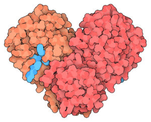
X-ray crystallographyThe main protease, resolved

This protease molecular structure was obtained from X-ray diffraction data by the team of Liu X., Zhang B., Jin Z., Yang H. and Rao Z. At the time it was still to be published, yet its early release in the PDB could aid the search for a vaccine.
Function flows from form, to a large extent, so such information is vital to understanding the new virus. Researchers can look for similarities to other protease structures from other viruses — which can suggest treatments that may work in similar cases — or be inspired to develop entirely new treatments.
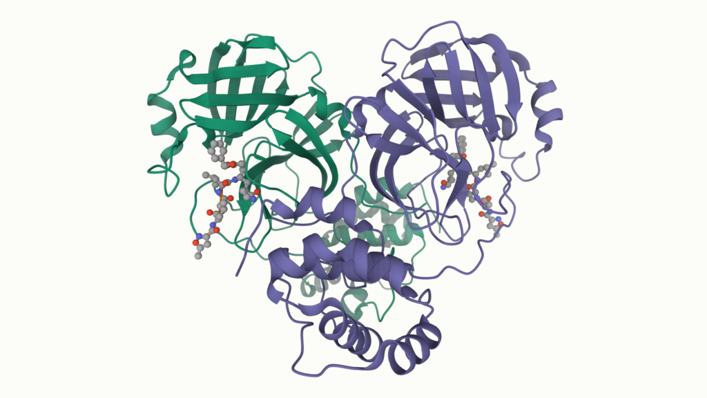
Cryo-EMA complementary view of the spike
An equally important source of structural information to X-rays is the complementary method of cryo-electron microscopy (cryo-EM). This new class of electron microscope can also reconstruct near-atomic-scale 3D images of large biomolecules. These facilities are sometimes made available at light sources using an access model similar to that of a regular beamline.
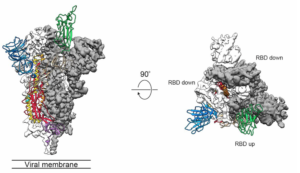
The protein spike of the coronavirus, shown here, participates in the attachment and infection process as the virus approaches and interacts with human cells. Its structure was obtained by Wrapp D., Wang N., Corbett K.S., Goldsmith J.A., Hsieh C-L., Abiona O., Graham B.S. and McLellan J.S., and was already published in Science at the time of writing.
Once again, an understanding of its structure and function can lead to the development of complex molecules that inhibit its function — thereby acting as a vaccine.
These are two examples of structural studies, and they spawn many “educated guesses” that ultimately lead to further studies, new knowledge, and finally the development of medical counter-measures. In due course, subjecting the virus to the scrutiny of science at such high precision will lead to a treatment, much as it has done in so many other cases. Let’s hope it can be fast.
Around the worldLight sources on the coronavirus front line
A quick scan of light-source activity around the coronavirus (as at 3 March 2020) reinforces the role these facilities play. Major synchrotrons had already contributed key structural studies of coronaviruses:
- ESRF — a structural view of coronavirus cell entry and neutralisation.
- APS — research aimed at stopping the coronavirus, and the discovery of a new coronavirus protein that reveals a drug target.
- SSRF — a pan-coronavirus fusion inhibitor targeting the HR1 domain of the human coronavirus spike.
- BSRF — the structure of the main protease from human coronavirus NL63, offering insights for wide-spectrum anti-coronavirus drug design.
- PETRA III — structural characterisation of the human coronavirus NL63 N protein.
Light sources declared an essential service
As the crisis deepened, several facilities were recognised as an essential service and remained open specifically for research to fight the coronavirus. NSLS-II, for example, offered a streamlined and expedited rapid-access proposal process for groups needing beam time for COVID-19 structural-biology projects. Its Center for Biomolecular Structure team supported remote macromolecular crystallography experiments at beamlines 17-ID-1 (AMX) and 17-ID-2 (FMX). The hope, expressed at the time, was that policymakers and the public would learn that we must invest in top-quality human-capacity development and significant facilities — within a culture of generating new knowledge for the common good.
A running logHow the effort unfolded
So much was happening that not all of it could be recorded, but the timeline below captures the milestones tracked on this page as the global structural-biology community mobilised against COVID-19.
- 2 Apr 2020
A dedicated database of the structural-biology effort from light sources around the world was launched.
- 7 Mar 2020
Light sources increasingly recognised as an essential service, with several remaining open for coronavirus research.
- 8 Mar 2020
European facilities (open to the world) coordinate their response: photon sources via the LEAPS initiative and neutron sources via the LENS initiative. A weekly #LightSourceScience and #SARSCoV2 research bulletin begins.
- 9 Mar 2020
The IUCr Newsletter (2020, 28/1) publishes an editorial: “Visualizing an unseen enemy — mobilizing structural biology to counter COVID-19.”
- 11 Mar 2020
A structure-based study of the antiviral drug Remdesivir provides critical insights into the working mechanism of viral RNA replication and a rational template for drug design to combat the infection.
- 11 Mar 2020
South African work sequences the complete SARS-CoV-2 genome from a local COVID-19 patient, showing how next-generation sequencing of pathogens can offer new insights into disease transmission and aid drug and vaccine design.
- 11 Mar 2020
A summary of ACE2 as a rational frontline therapy for COVID-19 — also a South African research focus, with work showing that ACE-domain selectivity extends beyond the directly interacting residues at the active site.
- 19 Mar 2020
Brookhaven Lab mobilises resources against COVID-19, combining expertise across disciplines to address drug development, medical supplies and information processing.
- 12 May 2020
A LEAPS review — Europe’s accelerator-based photon sources join forces in fighting COVID-19.
- 10 Jun 2020
An IUCr cover article introduces crystallography research on SARS-CoV-2 via X-ray diffraction at synchrotrons around the world.
- 26 Sep 2020
A CERN Courier feature: “Synchrotrons on the coronavirus frontline.”
- 16 Nov 2021
The Swiss Light Source (PSI/SLS) publishes a compendium of its COVID-19 research results.
Why this matters for Africa
The same structural science that let the world map the coronavirus within weeks is exactly the capability an African Light Source would bring to the continent — high-quality human-capacity development and major facilities, sustained by a culture of generating new knowledge for the common good.